Label the transmission electron micrograph of the cell: Unveiling the intricate world of cellular structures through the lens of advanced microscopy. This guide provides a comprehensive overview of the principles, applications, and techniques involved in labeling and interpreting transmission electron micrographs, empowering researchers and students to decipher the secrets hidden within the ultrastructure of cells.
Transmission electron microscopy (TEM) offers an unparalleled glimpse into the cellular realm, revealing the intricate details of organelles, membranes, and cytoskeletal components. By labeling these structures, we gain invaluable insights into their functions, interactions, and the overall organization of the cell.
Transmission Electron Microscopy (TEM)

TEM is a powerful imaging technique that uses a beam of electrons to create high-resolution images of biological structures at the nanoscale. It is widely used in cell biology to study the ultrastructure of cells and their organelles.
The principle of TEM is based on the interaction of electrons with matter. When a beam of electrons passes through a specimen, some electrons are absorbed, scattered, or deflected. The resulting pattern of transmitted electrons contains information about the structure and composition of the specimen.
TEM is used in a wide variety of applications in cell biology, including:
- Studying the structure and organization of organelles
- Investigating the dynamics of cellular processes
- Identifying and characterizing viruses and other pathogens
- Developing new drugs and therapies
To prepare a specimen for TEM imaging, it must be fixed, dehydrated, and embedded in a resin. The specimen is then cut into ultrathin sections and stained to enhance contrast.
A TEM setup consists of several key components:
- Electron gun: Emits a beam of electrons
- Condenser lenses: Focus the electron beam on the specimen
- Specimen stage: Holds the specimen in place
- Objective lenses: Magnify the transmitted electrons
- Image detector: Converts the magnified electrons into an image
Cell Structure and Organelles

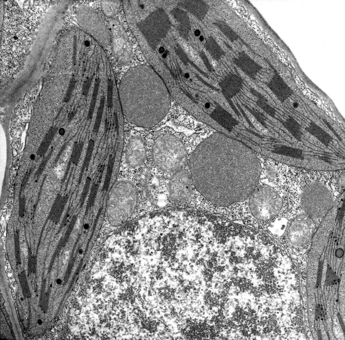
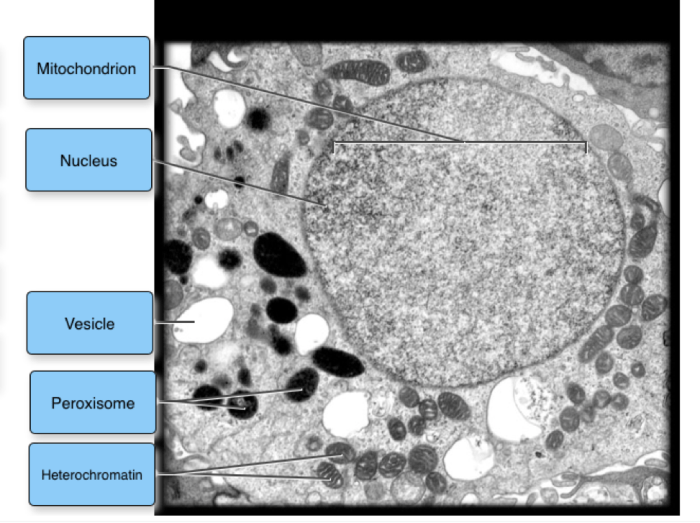
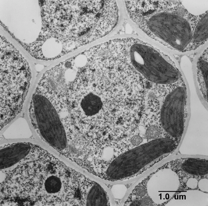
TEM micrographs provide detailed images of the ultrastructure of cells and their organelles. The following table identifies and labels the major organelles visible in a TEM micrograph of a cell:
| Name | Function | Size (nm) |
|---|---|---|
| Nucleus | Contains the cell’s genetic material | 10,000-15,000 |
| Mitochondria | Produce energy for the cell | 500-1,000 |
| Endoplasmic reticulum | Synthesizes and transports proteins | 50-100 |
| Golgi apparatus | Modifies and packages proteins | 50-100 |
| Lysosomes | Digest cellular waste | 50-100 |
| Peroxisomes | Detoxify harmful substances | 50-100 |
Each organelle plays a specific role in cellular processes, contributing to the overall function and survival of the cell.
Cell Membrane and Cytosol

The cell membrane is a thin, flexible barrier that surrounds the cell and regulates the exchange of substances between the cell and its environment.
In TEM micrographs, the cell membrane appears as a tripartite structure consisting of:
- Lipid bilayer: A double layer of phospholipids
- Transmembrane proteins: Proteins that span the lipid bilayer
- Glycocalyx: A layer of carbohydrates attached to the transmembrane proteins
The cell membrane plays a crucial role in maintaining cell integrity, protecting the cell from its surroundings, and regulating the transport of nutrients and waste products.
Cytoskeleton and Cell Shape

The cytoskeleton is a network of protein filaments that provides structural support for the cell and plays a role in cell division and intracellular transport.
In TEM micrographs, the cytoskeleton appears as a complex network of filaments. The three main types of cytoskeletal filaments are:
- Microtubules: Thick, hollow filaments
- Microfilaments: Thin, solid filaments
- Intermediate filaments: Filaments of intermediate thickness
Each type of cytoskeletal filament has a specific composition and function.
| Type | Composition | Role |
|---|---|---|
| Microtubules | Tubulin | Maintain cell shape, intracellular transport, cell division |
| Microfilaments | Actin | Cell movement, cell shape, cell division |
| Intermediate filaments | Keratin, vimentin, desmin | Provide structural support, maintain cell shape |
The cytoskeleton is essential for maintaining cell shape, organizing cellular processes, and enabling cell movement.
FAQ Section: Label The Transmission Electron Micrograph Of The Cell
What is the principle behind transmission electron microscopy (TEM)?
TEM utilizes a focused beam of high-energy electrons to penetrate and interact with a thin biological specimen, generating detailed images of its internal structures.
How are cells prepared for TEM imaging?
Cells are typically fixed, dehydrated, and embedded in a resin to preserve their structure and enhance contrast for electron imaging.
What are the key organelles visible in a TEM micrograph of a cell?
TEM micrographs reveal organelles such as the nucleus, mitochondria, endoplasmic reticulum, Golgi apparatus, lysosomes, and peroxisomes, each with distinct morphologies and functions.

